Understanding Osgood Schlatter Disease and Severs Disease
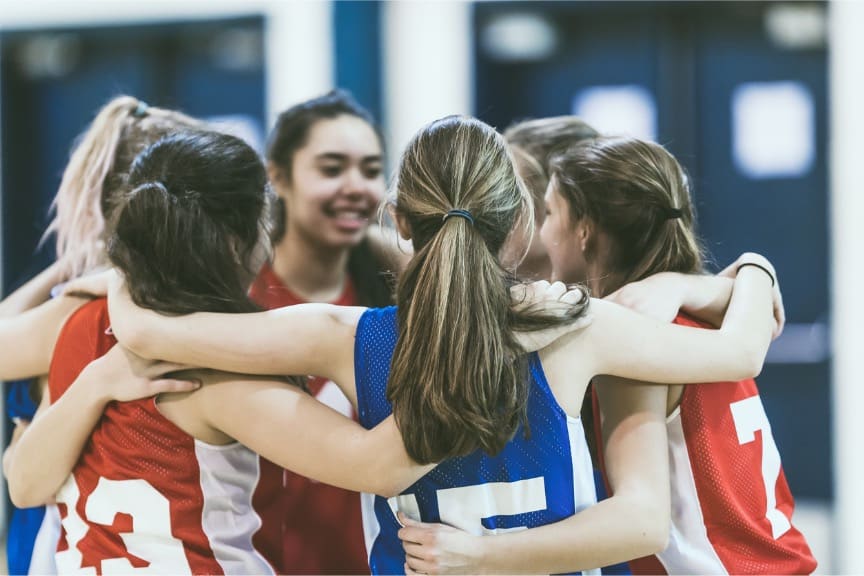
In the spring we see these two typical tween/teen painful diseases pop up prolifically because spring sports are in full gear! But, good news is on the way – they do grow out of it.
Let’s begin with Osgood Schlatter disease or Osgood Schlatter lesion: a very common cause of knee pain in children between the ages of 10 and 15 years old. It was named after two physicians in 1903, Dr. Robert Osgood and Dr. Carl Schlatter. Early diagnosis and appropriate treatment, especially rest, is essential as this injury can be stubborn to treat if left untreated. Please see your Doctor or Orthopedist at this point, or any point of pain to rule anything else out.
Osgood Schlatter disease symptoms
Osgood Schlatter knee pain is usually localized to a specific point at the tibial tuberosity and is therefore fairly easy to diagnose by a doctor. Tenderness and pain will be felt at the tibial tuberosity and will get worse during exercise, particularly weight-bearing or impact type exercise such as running, football, jumping, basketball, and similar sports. Pain will also be worse after exercise.
The athlete is likely to experience pain when contracting (tensing) the quadriceps muscles when the leg is straight or performing squat exercises. In particular, a single leg squat is likely to reproduce symptoms.
The tibial tuberosity may become swollen or inflamed and may even become more prominent than normal. There is likely to be localized swelling over the lower front of the knee and inflammation of redness may be seen. If the athlete has suffered for some time, then they will likely have atrophy (wasting away) of the quadriceps of thigh muscles. In extremely severe cases they (doctors) may do an x-ray to see exactly how much damage has occurred.
It is possible for Osgood Schlatter disease symptoms to come and go, becoming more or less severe, sometimes for no reason. It is a good idea to keep a training diary monitor training activity and symptoms to identify if any training methods worsen the injury.
Over time it can become progressively worse as tendon pulls at the growth plate at the top of the tibia. With repeated trauma new bone grows back as part of the healing process which causes an obvious prominent bony lump felt at the tibial tuberosity.
As the young athlete’s bones grow quickly, it can take some time for the muscles and tendons to catch up. If the muscles have not yet adapted to the length of the bones, then this can result in additional strain being placed at the point where the muscle attaches to the bone (via the tendon). This is frequent in younger people because their bones are still soft and are not yet fully grown.
Osgood Schlatter syndrome is primarily an overuse injury although certain factors can increase the likelihood of sustaining this condition.
Age – It is more likely to affect boys aged around 13 to 15 years old than girls, although girls certainly can be affected and if they are it is more likely to occur earlier at about aged 10 to 12 years old. it is often put down to growing pains in knees.
Activity – As the young athlete’s bones grow quickly, it can take some time for the muscles and tendons to catch up. These changes result in a pulling force from the patella tendon, on to the tibial tuberosity at the top of the shin. This area then becomes inflamed, painful and swollen
Osgood Schlatter Treatment
Treatment for Osgood Schlatters disease according to the experts consists of reducing pain and inflammation by applying the PRICE principles of protection, rest, ice, compression and elevation along with longer-term managing the condition through training modification and educating the athlete or parent until the young athlete grows out of it.
Once normal daily activities are pain-free then gentle stretching exercises may be beneficial along with sports massage for the quadriceps and myofascial release techniques to help stretch the muscles to ensure they are strong enough to cope with the loads placed on them as well as not being too tight. Massage should not be applied directly to the tibial tuberosity where the patella tendon inserts as this are likely to make symptoms worse. We have also had huge success with @rocktape kinesiology tapping for these issues.
Severs Disease
Sever’s disease mainly affects active children aged 8 to 15 years old and causes pain at the back of the heel. Overuse is often a contributing factor, but if managed correctly, it is something the young athlete should grow out of. Rest is an essential part of treatment, along with ice or cold therapy and managing training loads.
Sever’s disease symptoms
The main symptom of Sever’s disease is pain and tenderness at the back of the heel which is made worse with physical activity. It affects adolescent children who are often involved in a lot of sports training and physical activity. Tenderness will be felt, especially if you press in or give the back of the heel a squeeze from the sides.
There may be a lump over the painful area which develops over time. The pain may go away after a period of rest from sporting activities only to return when the young person goes back to training. Another common sign tight calf muscles which result in a reduced range of motion at the ankle. If the calf muscles are tight, then this increases the traction forces at the back of the heel.
Causes & anatomy
Sever’s disease is often associated with a rapid growth spurt. As the bones get longer, the muscles and tendons become tighter as they cannot keep up with the rate of bone growth. Tight calf muscles reduce the range of motion (dorsiflexion) at the ankle, resulting in more strain on the Achilles tendon and its attachment to the calcaneus or heel bone.
Sever’s Disease treatment
The aim of treatment is to reduce the pain and inflammation at the back of the heel. There is likely to be no magic instant cure and the young athlete may have to be patient while they grow. However, managing training loads to enable the young athlete to continue to get the most of out their sport is essential.
Initial treatment
Apply the PRICE principles of protection, rest, ice, compression, and elevation. Rest from any activity which makes the injury worse. Initially, certainly for the first 48 to 72 hours, this will a mean complete rest. If you continue to train on it when it is inflamed and painful, you will make it worse and possibly even cause more permanent damage to the bone.
Apply ice or cold therapy for 10 mins every hour initially, reducing the frequency as symptoms improve. Do not apply ice directly to the skin unless it is in the form of ice massage. Ice massage involves massaging an ice cube over the site of pain, ensuring the ice is not kept continually in once place. Ice can also be wrapped in a wet tea towel or better still, use a cold therapy and compression wrap. In the office we use @rocktape to tape this area which we have also seen amazing results with this issue.
Sports massage to the calf muscles may be beneficial in reducing any tension and helping the muscle to stretch. Massage directly to the site of pain at the back of the heel should not be done. This will only make the injury worse.
Results may not be evident for a few days or even weeks. It is also important to maintain a stretching/mobility/stability routine even after the injury has healed.
If this means training or playing slightly less to get ahead of the inflammation, then so be it. Better give it time to heal, then risk the possibilities of a major injury. Applying ice or cold therapy to the heel on a regular basis after training or exercise, even if it is not painful, may help reduce or prevent any pain and inflammation before it develops. We also suggest finding an experienced trainer who is certified in corrective movement, pt and additionally try acupuncture and cupping for best results, and please feel free to call us if you have any questions regarding how sports massage can help.
References
· Ogden JA, Ganey TM, Hill JD et al. Sever’s injury: a stress fracture of the immature calcaneal metaphysis. J Pediatr Orthop 2004;24:488-93
· Ramponi DR, Baker C. Sever’s Disease (Calcaneal Apophysitis) Adv Emerg Nurs J. 2019 Jan/Mar;41(1):10-14
· Brukner & Khan, “Clinical Sports Medicine”, 2012, McGraw Hill
· Knight, Kenneth L. “Cryotherapy in Sports Injury Management”, 1995, Human Kinetics
· Thompson & Floyd, “Manual of Structural Kinesiology”, 2001, McGraw Hill
· Denegar, Craig R. “Therapeutic Modalities For Muscular Skeletal Injuries”, 2006, Human Kinetics
· Kent, Michael. “Oxford Dictionary of Sports Science & Medicine”, 2003, Oxford University Press
· Reiman, Michael P. & Manske, Robert C. “Functional Testing In Human Performance” 2009, Human Kinetics
· Gallaspy, James B. & J.Douglas May. “Signs & Symptoms Of Athletic Injuries”, 1996, Mosby





Leave A Comment